Imaging Services

Service available at   Kapiolani Pali Momi Straub Benioff Wilcox

Imaging is an important diagnostic tool that is used by many medical specialties.

Our imaging staff is expertly trained and certified to provide you with accurate, efficient imaging services in a comfortable, supportive environment. 

Programs & Services available at:


Kapiolani 808 983 8626

Kapiolani specializes in diagnostic imaging for newborns, children, and teens.

That means everything, from its equipment to its pediatric protocols, are tailor-made for kids. Kapiolani uses the lowest radiation level of any CT scanner in the state. In fact, its levels are far below the American College of Radiology (ACR) recommendations.

MRI Information for Parents (PDF)

Kid Friendly CT Information for Parents (PDF)

Kapiolani Imaging: 1-808-983-8626

Pali Momi 808 535 7733

Pali Momi has been instrumental in elevating the level of care for Central and West Oahu.

It has pioneered health care for the communities it serves through investments in state-of-the-art technology. Pali Momi is home to West Oahu’s first permanent MRI, 320-slice CT scanner and cardiac catheterization program.

Pali Momi’s imaging department offers a full complement of comprehensive diagnostic imaging services and is equipped with the latest, most advanced technology. Pali Momi’s tradition of high quality care underscores its emphasis on ensuring the safety of its patients. The medical center’s safe scanning procedures ensure that the radiation dosage used in its procedures is far below the nationally recommended levels.

Additionally, Pali Momi offers convenient scheduling options including early mornings, evenings and weekends. Services include:

  • Computerized Tomography (CT) – featuring the new 320-slice CT scanner
  • Diagnostic – general radiology (available at Pali Momi Outpatient Center)
  • Electroencephalogram (EEG)
  • Mammography
  • Magnetic Resonance Imaging (MRI) – featuring our new 3T MRI, the highest-field strength available
  • Nuclear medicine studies
  • Osteoporosis (DEXA)
  • Special procedures in a newly renovated, comfortable space
  • Ultrasound – general, gynecological, obstetric and vascular

Making CT Scans Safer (PDF)

For appointments, call: 1-808-535-7733

Straub Benioff 808 522 4220

Straub offers a wide range of services, ranging from simple x-rays to complicated interventional radiological procedures.

We offer MRI (1.5T), helical CT scanners with low dose radiation, ultrasound, digital mammography and tomosynthesis*, nuclear medicine, and comprehensive angiography.

Straub also has the only upright stereotactic breast biopsy system in the state, which allows patients to undergo their breast biopsy in the sitting position, rather than lying face down. Digital mammography and X-ray are also available at most satellite clinic locations.

Straub’s staff is available to perform a wide variety of vascular and non-vascular interventional procedures, including: diagnostic angiography, venous evaluation of pelvic congestion syndrome, and embolization of uterine fibroids.

On staff are ten radiologists that are fellowship trained in the following subspecialties: neuroradiology (brain and spine), musculoskeletal (joints), cardiac (heart), mammography (breast), interventional radiology (imaging-guided procedures and treatment), and body imaging.

*Available only at Straub's King Street location

Straub Imaging Services: 808-522-4221


Wilcox 808 245 1030
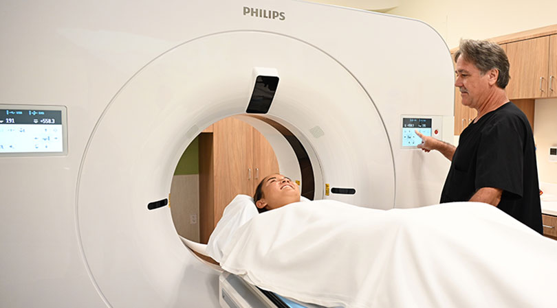
patient receiving a CT scan

The center is equipped with state-of-the-art technology to serve patients across the inpatient, outpatient and emergency settings.

The spectral-detector CT scanner produces comprehensive images for patients of all ages, including children, and with a range of medical needs, such as bariatrics. The machine generates high-quality, exceptionally clear images with faster setup times and shorter exam times. This results in a lower dose of radiation as well as a more comfortable overall experience.

The 320-slice CT scanner offers advanced imagery full-body scanning in a single rotation of the X-ray tube. This faster scan time is helpful for patients who have a hard time staying still for extended periods.

Advanced features of the CT scanners at Wilcox help medical teams detect, diagnose and monitor both common and chronic diseases, disorders and conditions, including cancer, heart disease and bone fractures.

Imaging services are available 24 hours a day, seven days a week. Board-certified physician radiologists provide quality care and timely interpretation of radiographic procedures.

Services are by referral from a patient’s primary care physician, who will use these internal images of the body to assist with diagnosis and treatment.

Wilcox Family Imaging Center: 1-808-245-1030

Useful Forms [PDFs]:

FAQs

Angiography

What is angiography?
Angiography is an x-ray examination of the blood vessels that allows doctors to see how blood circulates within the body. It is used when a blockage in the flow of blood or abnormality of a blood vessel is suspected.

An angiogram can tell a doctor whether an artery is blocked, where the blockage is, how severe it is and what the cause is. A common cause of blockage is a blood clot in an artery narrowed by arteriosclerosis, or hardening of the arteries.

How does an angiogram work?
A radiologist will place a thin, flexible tube called a catheter into an artery or vein and then inject a small amount of contrast dye into the blood vessel to make it more visible in an X-ray.

What is the angiogram process?
There are just a few steps in this simple and nearly painless procedure:

  • Patients are positioned on a comfortable table and X-rays are taken to get a pre-test picture.
  • Patients are then given a local anesthetic where the catheter will be inserted. A small incision will be made and a thin catheter inserted into the artery.
  • When the tube is exactly in place, some distance along the artery, the X-ray dye will be injected and the catheter removed. While the contrast dye is being injected, patients may experience a feeling of warmth, a metallic taste, or nausea. These symptoms are normal and should only last a minute or so.
  • Removal of the catheter tube does not hurt, but pressure will be applied when it is removed to prevent the artery or vein from bleeding.

The advanced X-ray camera used in our angiography testing will show exactly where the dye reaches and where it doesn't; indicating if there is a blockage or abnormality in the blood vessel.

The entire process usually takes one to two hours to complete.

How do I prepare for my angiogram?
Patients receive a call to discuss what they can and cannot do before and after their test. However, as a general guide:

  • Patients who are allergic to X-ray dye or iodine should let their doctor know as soon as possible.
  • Smokers should not smoke for at least 24 hours before their angiogram as nicotine may affect the results of some tests.
  • Patients who need to take medications in the morning should wash them down with just a few sips of water. They should also bring all medications with them to the hospital.
  • Patients should not eat any solid food after midnight the night before their procedure. Patients may, however, have clear fluids, such as water or broth.

What happens after the test?
If a patient is having the procedure done as an outpatient, they will remain in the recovery room for four to six hours following your angiogram. Although they will be asked to lie still, they will be able to eat and drink. Visitors are allowed.

Someone will need to drive the patient home, and the patient should not drive for the rest of the day. Patients should also relax and take it easy at home. If they have a desk job, they should be able to return to work the next day but should avoid any lifting or straining for at least a week.

Before the patient goes home, the radiology nurse will give them further instructions.

When will I get the results?
A radiologist will read the scan and send a report to the physician within 24 hours. The patient’s doctor will then contact them to discuss their results.

Will my insurance cover angiography?
Most insurance plans cover this test. Patients should contact their insurance plan if they have any questions.


CT Scan

Advanced imaging technology can better detect diseases at an early stage, when a wider array of effective treatment options may be available.

Faster scanning time with the 320-slice CT scanner

  • Technology is two to 10 times faster than most other CT scanners
  • 320 images can be acquired rapidly, reducing what used to take minutes down to seconds
  • Faster scanning time benefits patients, especially those with breathing difficulties or some other distress
  • Speed and detail aid in decision-making in emergency cases

3D images provide enhanced diagnostic capability
Detailed information captured by the 320-slice CT scanner is used to generate 3D images with greater anatomical detail.

How is CT scan used?
CT scans provide detailed views of many types of tissue, including the lungs, bones, soft tissues and blood vessels and allow doctors to more effectively detect and treat a range of life-threatening illnesses, such as cancer and lung disease.

The unmatched speed and detail of the new 320-slice CT scanner also aids in decision-making in emergency cases where physicians have to make critical decisions quickly.

Diagnosis made with the assistance of CT can eliminate the need for invasive exploratory surgery and surgical biopsy and is effective in determining, at an early stage, the appropriate treatment for patients.

What happens during the scan?
During the non-invasive test, the patient is placed on a table and moved incrementally through the squared off donut-shaped scanner while an X-ray beam is projected through cross sections of their anatomy. The X-ray energy passes through the patient and is recorded on electronic detectors in the scanner. This information is then sent to a specialized computer that reconstructs the information into individual slices and combines them sequentially into a comprehensive volume image of the entire scanned area. The thinner the slices, the more revealing the detail is in the resulting images and the more definitive the exam results.

When will I get the results?
A radiologist will read a patient’s scan and send a report to their physician within 24 hours. Their doctor will contact them to discuss their results.

Will insurance cover a CT scan?
Most insurance plans cover this test. Patients should contact their insurance plan if they have any questions.


MRI

Will the scan hurt?
No, patients won't feel a thing. They simply lie still during the scan.

Is it safe?
MRI technology has been available for more than 20 years and is extremely safe. Unlike X-rays, MRI does not use radiation.

How long does it take?
Depending on each situation, an MRI could take between 30 and 60 minutes.

What happens during the scan?
It's really very simple.

  • Patients are positioned on the scanning bed.
  • An instrument called an image coil will be placed over the area examined.
  • The bed (tumbrel) will then be moved into the scanning chamber.
  • Patients will be asked to lie very still during the scan.
  • A doctor may request that a patient receive an injection of contrast agent to provide additional information about the area being scanned. The injection feels like any other shot, and is very safe.
  • During the scan, patients may hear a chirping sound from the scanner. This is normal.

Can anyone have an MRI?
Since MRI is very safe, most people can have a scan done. However, if a patient has a pacemaker or metal implant, or if they are a woman in their first trimester of pregnancy, they cannot be scanned.

When will patients get the results?
A radiologist will read a patient’s scan and send a report to their physician within 24 hours. Their doctor will contact them to discuss their results.

Will insurance cover an MRI?
Most insurance plans cover MRI. Patients should contact their insurance plan if they have any questions.

How should a patient prepare for their scan?

  • Patients should arrive 30 minutes before their scheduled appointment and go to the Registration Department.
  • Patients should dress in warm, comfortable clothing (sweatshirt and pants work well).
  • Patients cannot wear anything metallic during the exam, so it's best for them to leave watches, jewelry or anything else containing metal at home.
  • If sedation is ordered, a doctor will provide further instructions.

%MCEPASTEBIN%